Medical professionals could use the technology to reach parts of the body that are hard to get to by making small cuts in the skin or using natural openings.
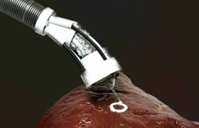
UNSW Sydney engineers have created a small and flexible soft robotic arm that could be employed to 3D print biomaterial direct onto organs within a person’s body.
3D bioprinting is the method of creating biomedical components out of bioink to create real tissue-like structures.
Bioprinting is mostly used for research, like tissue engineering and making new drugs. To make cellular structures outside of a living body, scientists usually use large 3D printing machines.
In a paper published in Advanced Science, researchers from the UNSW Medical Robotics Lab, led by Dr. Thanh Nho Do and his PhD student, Mai Thanh Thai, as well as Scientia Professor Nigel Lovell, Dr. Hoang-Phuong Phan, and Associate Professor Jelena Rnjak-Kovacina, describe their new research.
The result of their work is a small, flexible 3D bioprinter that can be put into the body like an endoscope and deliver multilayered biomaterials directly to the surface of organs and tissues inside the body.
The F3DB prototype is a remotely operated, long, flexible robotic arm with a swiveling head that “prints” the bioink.
According to the study team, with additional development and maybe within five to seven years, medical experts might employ the technology to access difficult-to-reach places within the body via minute skin incisions or natural orifices.
The device has a printing head with three axes that is directly attached to the end of a soft robotic arm. Its printing head functions very similarly to other desktop 3D printers since it is made of soft artificial muscles that enable it to move in three dimensions.
Due to hydraulics, the soft robotic arm can bend and twist and may be made to any necessary length. Its stiffness may be precisely adjusted by using various elastic fabric and tube kinds.
When more sophisticated or arbitrary bioprinting is needed, the printing nozzle may be manually manipulated or programmed to print certain forms. The researchers also used a controller that is based on machine learning and can help with printing.
The UNSW group also conducted cell viability tests on live biomaterial produced using their technique to prove the technology’s viability.
The majority of the cells were found to remain alive after printing, demonstrating that the cells were not harmed by the technique. After seven days of growth, the cells doubled in size, and four times as many cells were still visible one week following printing.
The study team also showed how the F3DB may be utilized as an all-purpose endoscopic surgical instrument to carry out a variety of tasks.
According to them, endoscopic submucosal dissection (ESD), a procedure used in surgery to remove some tumors, particularly colorectal cancer, might be particularly crucial in this situation.
Colorectal cancer is the third most common type of cancer death in the world. However, if colorectal neoplasia is removed early, the patient’s chance of living for five years goes up by at least 90%.
To label and subsequently remove malignant tumors, the F3DB printing head’s nozzle may be used as a kind of electric scalpel.
Water can also be sent through the nozzle to clean up blood and extra tissue at the site, and biomaterial can be printed in 3D right away while the robotic arm is still in place to speed up the healing process.
It was shown that the F3DB is capable of performing such multi-functional operations on a pig’s intestine, and the researchers claim that the findings indicate that the F3DB is a good candidate for the creation of an all-in-one endoscopic surgical instrument in the future.
“Compared to the existing endoscopic surgical tools, the developed F3DB was designed as an all-in-one endoscopic tool that avoids the use of changeable tools which are normally associated with longer procedural time and infection risks,” adds Mai Thanh Thai.
The next step for the system, which has been given a provisional patent, is to test it on living animals to show how it works in the real world.
The team also intends to install further features, such as a real-time scanning system and integrated camera, to recreate 3D tomography of living tissue.
