

Cochlear implantation is a well-established means to treat severe hearing loss has been linked to new bone formation, increased Hearing Loss.
A cochlear implants may adversely affect long-term hearing preservation, says new study.
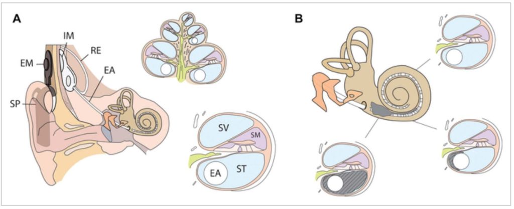
Cochlear implantation is a time-honored method of treating severe hearing loss. The device is composed of two components: an external half that is worn behind the ear and a surgically implanted second part that stimulates nerves in the cochlea, a fluid-filled spiral structure in the inner ear that conveys sound to the brain via sensory neurons. While cochlear implants can not restore normal hearing, they can assist a person in recognizing words and comprehending speech.
While cochlear implant side effects or issues are uncommon, post-mortem research has connected them to inflammation, fibrosis, and new bone development. The possible clinical consequences of new bone formation make it desirable to see and maybe block development, yet in-vivo—or within the body—detection has not yet been demonstrated.
“Such subtle changes are challenging to visualize in vivo, in particular in the vicinity of a metallic implant causing artifacts on computed tomography images,” says study co-lead author Dr. Floris Heutink.
“However, through the new availability of an ultra-high-resolution CT scanner, we were encouraged to investigate this.”
Dr. Heutink and colleagues assessed new bone development and its implications in 123 patients with cochlear implants using ultra-high spatial resolution CT (UHRCT).

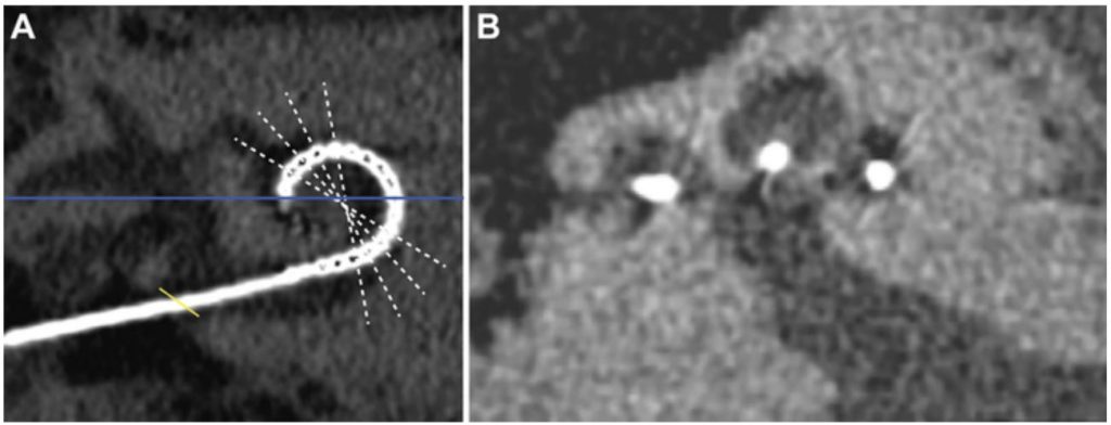
Using UHRCT, the researchers were able to identify new bone development in vivo. Within four years of implantation, 83 (68%) of the 123 patients had new bone development, primarily at the base of the cochlea. The group with new bone growth had a much greater long-term residual hearing loss.
“As indicated by our study, there is a correlation between new bone formation and long-term residual hearing loss,” adds study co-author Dr. Berit M. Verbist.
The growth of new bone around the cochlear implant electrode has a number of unfavorable repercussions due to the device’s and the surrounding structures’ impact. It may impair the propagation of electrical current within the cochlea, resulting in more complicated device fitting, channel interaction, and a less satisfactory overall hearing outcome. Additionally, it may complicate future therapies such as gene therapy for cochlear restoration.


“Last but not least, new bone formation may complicate reimplantation surgery,” says Dr. Verbist.

The researchers stated that advancements in design and surgical techniques have enabled an increasing number of individuals with severe to profound hearing loss to have cochlear implantation. This increase in patients underscores the critical nature of developing a mechanism for identifying and monitoring new bone growth in vivo. Dr. Heutink noted that numerous unanswered concerns must be addressed before detecting new bone development in an individual patient would have therapeutic implications.
“This technique will be a valuable tool to gain insight into occurrence, time course and the pathophysiology of this process and maybe used to evaluate still-to-be-developed treatments against new bone formation,” he concluded.
Source: Radiological Society of North America
Image Credit: iStock and Radiological Society of North America
You were reading: CT Reveals New Bone Formation after Cochlear Implant


