

The electron microscope image of Omicron strain
The virus is represented by clusters of characteristic spherical objects with crown-shaped spikes on their surface.
Scientists from the University of Hong Kong have photographed the Omicron coronavirus strain.
It is noted that the researchers were the first to make an electron micrograph of a monkey kidney cell (Vero E6) that was infected with Omicron.
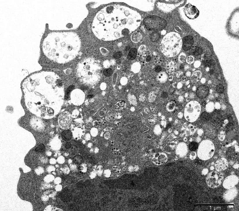
The picture shows lesions with swollen vesicles, which contain viral particles. Scientists also managed to make out clusters of characteristic spherical objects with crown-shaped spikes on their surface.

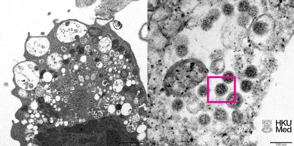
(Left) Low magnification electron micrograph of a monkey kidney cell (Vero E6) after infection with the SARS-CoV-2 Omicron variant showing cell damage with swollen vesicles containing small black viral particles.
(Right) High magnification electron micrograph of an infected Vero E6 cell showing aggregates of viral particles with corona shaped spikes on their surface (red box).

Source: HKUMED
Image Credit: Getty
You were reading: This is how Omicron variant look-like


