
Early-Stage Alzheimer’s: A new study reveals how cognitive decline begins and progresses following amyloid build-Up.
Using a novel brain imaging technique, neuroscientists at the Medical University of South Carolina have discovered subtle differences in the way the brains of older adults with preclinical Alzheimer’s disease (AD) function.
These findings, published in Brain Connectivity, suggest that individualized brain fingerprints could be used to uncover early signs of Alzheimer’s disease in patients who have no noticeable symptoms of cognitive decline but show early indications of the disease, such as the buildup of amyloid-beta proteins in their brains.
In an effort to study the preclinical phase of Alzheimer’s disease (AD), a research team led by Andreana Benitez, Ph.D., and Stephanie Fountain-Zaragoza, Ph.D., has developed a novel brain imaging analysis technique to construct individualized maps of brain function.
By analyzing the links between subtle changes in brain function and declining cognitive performance, assessed through behavior-based tests, the team has discovered a potential brain-based reason for very subtle cognitive changes in the early phase of AD.
This breakthrough could offer a new approach for studying the preclinical phase of the disease, where prior studies have not found an association between brain function and behavior.
Improved brain mapping has enabled the detection of subtle changes in brain function, which is crucial in studying the preclinical phase of Alzheimer’s disease (AD) to gain a deeper understanding of its onset and progression. Despite the challenges posed by the subtle nature of these changes, researchers at MUSC were able to identify them through a novel brain mapping technique that was funded by the South Carolina Clinical & Translational Research Institute. The technique, known as functional connectome, involves the measurement of how different brain regions communicate with each other, similar to the way neighborhoods in a city are connected by highways. By observing the activity across the brain using this technique, researchers were able to gain insights into the amount of activity within each brain region and how efficiently information is transmitted between them.
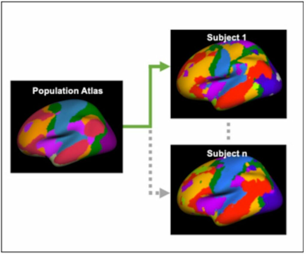
To better understand how brain regions function in individuals, the researchers utilized a more recent and highly sensitive form of image analysis. This technique, called the individualized functional connectome, was developed by their collaborator, Dr. Hesheng Liu. In contrast to traditional functional connectomes that use an average of many people’s brains as a map for functional brain regions, Liu’s method can reveal unique patterns of brain function for each individual.
According to Fountain-Zaragoza, while we all have the same functional parts of the brain, they are positioned slightly differently, akin to a fingerprint. This approach creates an individualized brain fingerprint that provides a more precise representation of the different functional regions in each person’s brain.
To investigate subtle changes in brain function, the researchers employed the innovative brain fingerprinting technique on a group of 149 participants aged between 45 to 85 who did not display any signs of cognitive decline. The participants underwent PET scans to assess for early amyloid-beta protein buildup and were divided into two groups based on the presence or absence of these biomarkers. Brain fingerprints were generated using MRI scans for all participants.
The researchers then evaluated the performance of each group on behavior-based tests of information processing. They observed that specific alterations in the brain fingerprint were linked to poorer information processing in those with preclinical AD or amyloid-beta buildup.
In those with preclinical AD, individuals with higher between-network connectivity, or more activity on the brain’s highways, showed worse information processing. Conversely, participants with higher within-network connectivity, or greater brain activity within important brain regions, demonstrated better information processing.
“A healthy brain typically has a balance of connectivity within and between its networks,” points out Fountain-Zaragoza.
They “found that in preclinical AD—when amyloid build-up is present in the brain—this balance can be disrupted, potentially leading to information no longer being processed as efficiently.”
The study’s findings indicate that the individualized functional connectome technique can identify subtle changes in brain function that may not be detectable with traditional brain imaging analysis methods.
Moreover, the research suggests that early amyloid-beta protein buildup can have an impact on brain network function, even prior to the onset of observable cognitive decline symptoms.
Furthermore, the study highlights that changes in connectivity within and between specific brain networks may indicate early issues with information processing. Addressing this connectivity imbalance could represent a promising target for therapies designed to enhance outcomes for individuals with AD.
Having secured a fresh round of grant funding from the National Institute on Aging, Benitez and Fountain-Zaragoza plan to move forward with their research on preclinical AD. Going forward, they intend to delve deeper into how brain changes impact disease progression and investigate the efficacy of new treatments, such as brain stimulation, that may be able to slow down AD.
“There’s a lot of great work aimed at helping us understand the earliest signs and symptoms of Alzheimer’s disease,” adds Fountain-Zaragoza. “This area of work is important for understanding the full spectrum of the disease and identifying who might be at risk of developing it.”
Source: 10.1089/brain.2022.0032
Image Credit: Getty